APPROVED
Digital retinal photos or OCT? Choosing the right imaging for your eyes
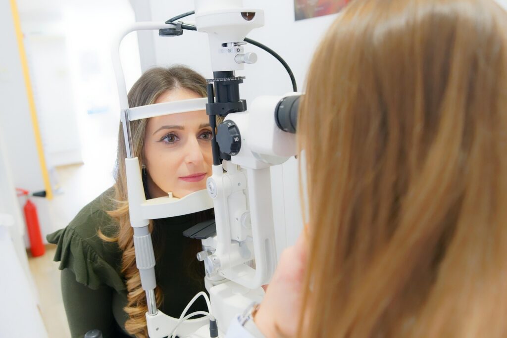
Advanced imaging has changed how we protect vision. Clear, detailed pictures of the retina let your doctor spot tiny changes before you notice symptoms, guide treatment, and compare year to year to keep you seeing your best.
At B&B Optometry in Yorba Linda and Corona, we use two main tools for retinal imaging: high-resolution digital retinal photos and Optical Coherence Tomography (OCT). Each test answers a different clinical question. Together, they help us detect problems early and track your eye health with confidence.
If you have questions about comfort, driving afterward, timing, or insurance, you are not alone. This guide explains what each test shows, when we recommend it for diabetes, macular degeneration, and glaucoma, and how it fits into your comprehensive exam.
What a digital retinal exam is and what it shows
A digital retinal exam uses a specialized camera to capture a color photograph of the back of your eye. The image shows the retina, macula, optic nerve, and blood vessels. It documents surface appearance, pigment patterns, tiny hemorrhages, drusen deposits, vessel caliber, and optic nerve contour.
- What you feel: a brief flash of light and a few seconds of stillness while the picture is taken.
- Time: typically seconds to a couple of minutes for both eyes.
- Dilation: at our Yorba Linda office, many patients can have no-dilation imaging with our iCare retinal camera. In some cases, dilation is still recommended for a wider or more detailed view.
Digital photos create a visual record. On follow-up visits, we place new images side by side with prior images to look for subtle change. That long-term comparison is one of the biggest advantages of retinal photography.
What OCT is and what it shows
Optical Coherence Tomography (OCT) is a non-contact scan that uses light waves to capture cross-sectional images of the retina and optic nerve. Think of it like ultrasound with light. Instead of a surface photo, OCT shows the layers inside the retina and the thickness of nerve tissue.
- What you feel: you look at a fixation target; there is no puff and no touch.
- Time: usually a few minutes per eye, including alignment and scan capture.
- Dilation: not always required. Your doctor will decide based on pupil size, media clarity, and the area we need to evaluate.
OCT helps detect fluid in or under the retina, swelling from diabetic macular edema, thinning of the retinal nerve fiber layer in glaucoma, and structural changes in macular degeneration that are not visible on a standard photo.
Digital photos versus OCT, in plain language
- The photo is a color snapshot of the retinal surface. It is excellent for documenting spots, pigment, hemorrhages, vessel changes, and optic nerve appearance.
- OCT is a cross-section. It is excellent for measuring thickness, identifying hidden fluid, mapping layers, and tracking microscopic change.
Your doctor may order one or both on the same day. They answer different questions and complement each other.
When we recommend each test
- Diabetes: Digital retinal photos help document microaneurysms, hemorrhages, and vessel changes. OCT helps detect and monitor diabetic macular edema. Many diabetic visits include both tests to capture surface findings and hidden swelling. If you are due for diabetic screening, our team can help you schedule through our page for diabetic eye tests in Corona or Yorba Linda diabetic eye screenings.
- Macular degeneration: Photos document drusen and pigment patterns. OCT reveals subtle fluid, atrophy, and layer integrity to guide treatment timing and referrals.
- Glaucoma: Photos provide a baseline of optic nerve appearance. OCT measures nerve fiber and ganglion cell complex thickness to catch small changes over time. We also pair these with eye pressure checks when risk is present.
If you are in the Corona area and need a digital retinal imaging exam, learn more about our medical eye care and imaging options on our ocular disease page.
How long testing takes and what to expect
Most imaging adds only a few minutes to your visit. Photos take seconds. OCT scans take a few minutes per eye. The team positions you comfortably, explains each step, and checks image quality before you leave so we do not need to repeat scans later. Results are reviewed with you the same day whenever possible. We save images to your chart for side-by-side comparison at future exams.
Can you drive afterward?
- Digital photos without dilation: most patients can drive and return to work or school right away.
- With dilation: your near vision and light sensitivity can be affected for a few hours. Many patients still drive safely with sunglasses, but comfort varies. If you are unsure, bring a driver or plan extra time before driving.
- OCT: the scan itself does not affect driving. If dilation is not used, you can typically drive immediately.
At Yorba Linda, our no-dilation option often makes it easier to continue your day. Your doctor will still recommend dilation when a wider view is needed.
Is digital retinal imaging worth it?
In our experience, yes. The value is cumulative. A single photograph is helpful, but the real power comes from comparisons over years. We can spot a new tiny hemorrhage, a subtle change in the optic nerve rim, or a slight shift in macular features that might signal early disease. Earlier detection often means simpler treatment and better protection of vision. Imaging also helps us explain findings clearly, which supports shared decision-making for your care.
Does insurance cover imaging?
Coverage varies by plan and by medical necessity. Many medical plans cover OCT and retinal photography when we are evaluating or monitoring a diagnosed condition, such as diabetes, glaucoma, or macular degeneration. Some vision plans contribute to screening photos during routine exams, while others consider them an out-of-pocket wellness upgrade. Our team checks benefits when possible and will discuss any expected costs before proceeding. Flexible Spending Account (FSA) or Health Savings Account (HSA) funds often apply to eligible out-of-pocket vision expenses.
Comfort, safety, and continuity across both locations
B&B Optometry provides full-scope, unhurried eye care in Yorba Linda and Corona with multilingual support in English, Spanish, and Mandarin. The same standards of clinical imaging, careful explanation, and follow-up apply in both offices. When diabetic changes, glaucoma risk, or macular findings are present, we coordinate with your primary care physician or a retinal specialist as needed and keep your imaging history organized so nothing gets lost between visits.
If you need guidance from an Optometrist in Yorba Linda or are ready to book eye exams in Corona, our team is here to help you choose the right tests and timing based on your history and goals.
FAQ: quick answers
- What is a digital retinal exam? A fast, high-resolution color photo of the back of the eye that documents the retina, macula, optic nerve, and blood vessels for today’s evaluation and future comparisons.
- What is the difference between OCT and digital retinal imaging? Photos show the surface in color; OCT shows cross-sectional layers and thickness to reveal hidden fluid or structural change. They are complementary, not interchangeable.
- Can you drive after digital retinal imaging? If no dilation is used, most people can drive right away. If you are dilated, expect light sensitivity and near blur for a few hours and consider arranging a ride.
- Is digital retinal imaging worth it? Yes, especially over time. Year-to-year comparisons help detect subtle change early and guide care.
- Does insurance cover digital retinal imaging? It depends on your plan and whether the test is medically necessary. We verify benefits when possible and review any expected costs with you before testing.
The bottom line
Digital retinal photos and OCT work together to protect your vision. Photos provide clear color documentation of the retinal surface, while OCT reveals cross-sectional detail that catches small changes early. Testing is quick, comfortable, and often does not require dilation in Yorba Linda. Over time, side-by-side comparisons add real value by showing what is stable and what needs attention. If you are due for a routine eye exam in Yorba Linda or want to discuss diabetic monitoring in Corona, contact B&B Optometry to schedule a visit. Our doctors will recommend the right imaging for your eyes and your peace of mind.